
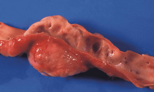
Esophageal Worm (Spirocerca lupi)
The esophageal worm is an underrated and potentially fatal spiral roundworm parasite in
domestic and wild dog breeds. Dogs become infected when they eat an intermediate host (dung beetle) or
a transport host (e.g. chicken viscera, reptiles, and rodents).
_ Parasite: Spirocerca lupi
_ Common name: Esophageal worm
_ Host: Dog breeds
– Incubation period: 5-6 months
_ Site of adult worms: Wall of the esophagus and stomach
_ Distribution: Tropical and subtropical regions
_ Route of transmission: Ingestion of an intermediate host or a secondary (transport) host
– Transmission to humans: No
_ Distribution
The esophageal worm is widespread across the tropical and subtropical regions of Asia, Oceania,
Latin America, Africa, and the Middle East.
_ Clinical signs
Dogs initially infected may show no symptoms, but the disease can progress to regurgitation, vomiting, black
tarry stools, and a gradual decline in health and weight loss due to granulomas in the esophagus and stomach.
Larvae migrating through the aorta can cause pleuritis, leading to coughing, dry retching, and difficulty
breathing. Aortic aneurysms can sometimes rupture, causing thoracic hemorrhage and sudden death. The
fibrous nodules in the esophagus and stomach can turn malignant and progress into connective-tissue cancer
in the esophagus with secondary metastatic tumors. Hypertrophic osteopathy with calcification of the
periosteum of the front legs is often found alongside chest-cavity lesions in dogs with S. lupi-associated
neoplasia.
_ Diagnosis
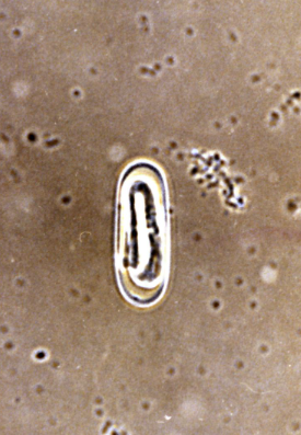
Eggs are shed intermittently in the stool, or not at all if the nodule has no fistula. Detecting
eggs with the characteristic elliptical, embryonated shape (small, 35 × 15 μm) in the stool via a standard
fecal flotation test using a solution with S.G. > 1.20 is the optimal diagnostic method. The
primary lesions seen on X-ray include a mediastinal mass, usually near the distal esophagus. Spondylitis
of the thoracic vertebrae is often seen on chest X-rays. Contrast
radiography and CT scanning are useful additional imaging methods. Esophagoscopy
has higher diagnostic sensitivity than X-ray.
BTM Pet Shop — Diagnosis and treatment for pets (24/7)
- ✂️ Pet grooming
- 🏨 Pet hotel
- 🎓 Grooming training for students
- 🛍️ Pet accessories supplied
- 📞 093 71 73 188
- 📍 135/31 Nguyễn Hữu Cảnh, Ward 22, Bình Thạnh, Ho Chi Minh City